New cancer scanner gives better quality for half the radiation

Particle physicists at the University of Oslo in Norway have created a new method of scanning for cancer that will provide a better image for doctors to examine and reduced radiation risk for the patient. Three different types of imaging technology exist: PET (Positron Emission Tomography), CT (Computerized Tomography), and MRI (Magnetic Resonance Imaging). Currently, the most common form of cancer examination involves comparing PET and CT scans.
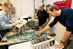
But scientists working on the project have combined PET with MRI, which does not emit radiation and provides a better image than CT. They did so by creating a PET scanner that was small enough to fit inside an MRI machine so that both types of scans can be captured at once. The new PET scanner developed also allows them to achieve better image quality, by placing it closer to the subject. Though the scanner was initially built for animals, researchers indicate that it could be used in hospitals in the future. According to researcher Erlen Bolle, "The high resolution in our PET scanner provides better images, and the high sensitivity makes it possible to use only half as much radioactivity in the examinations without it affecting the image quality."
[via ScienceDaily]
Related on SmartPlanet:
This post was originally published on Smartplanet.com