Building brains: toward a do-it-yourself guide

 With an estimated 100 billion neurons chattering to one another through perhaps a quadrillion synaptic connections, the human brain has the most intricately complicated structure and function of any object in the known universe. How do you build something like that? In fact, how does something so ferociously complex start off as a single cell and then create itself?
The problem might at first seem impossible to fathom, even with the knowledge that genetics, natural selection, and a half billion years of evolution can accomplish amazing things. But complexity does not have to be born of complexity, as mathematics and computer programs routinely demonstrate. Very simple equations can give rise to the stunning complexity of fractals, for example.
Several recent neuroscience studies may be illuminating some of the structural and development principles that organize the brain. Those principles include:
- Co-opting older, simpler genetic programs and elaborating on them to new effect.
- Maintaining geometric order.
- Relying on hierarchies of control.
True, science is still very far from knowing enough to define confidently how a brain takes shape (let alone how one works). Yet it's still fascinating to see how much structure may be determined by relatively simple rules of thumb.
Rule 1: Elaborate on older genetic mechanisms
In the earliest stages of embryonic development, the nervous systems of vertebrate animals are much like those of simpler invertebrates such as worms and starfish. The big differences start to appear as a result of actions by certain parts of the vertebrate embryo's body called signaling centers. These centers release proteins that tell portions of the embryo's exterior layer of tissue (the neuroectoderm) to organize themselves into major divisions of what will eventually become brain and spinal cord.
The standing inference has been that the genes for those signaling proteins emerged as part of whatever evolutionary changes first split the vertebrate and invertebrate lines. New work by Ariel M. Pani of the University of Chicago and others, recently published in Nature, suggests that is not the case, however.

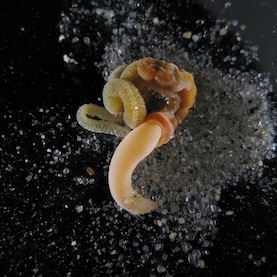
Pani and her colleagues have identified a highly similar set of genes active in the acorn worm (Saccoglossus kowalevskii), a tiny aquatic invertebrate. According to their studies, not only do these genes have sequences that echo those of the vertebrates but they also express themselves in a similar pattern and in corresponding parts of the body. They also help to organize the formation of features in the worms' ectodermal tissue layer.
What's most curious about Pani's findings -- and makes them controversial -- is that the acorn worms fall into the hemichordate branch of the invertebrate family, and as such are much more distant cousins to vertebrates than are many other creatures (such as sea squirts) that seem to lack these signaling center genes. The implication is that the genes were present in some common wormy ancestor from about 500 million years ago but that the other lines of related invertebrates subsequently lost them during evolution. Yet some biologists question both whether the genes are truly missing from the other invertebrates and whether they really serve to organize the acorn worms' ectoderm to the sophisticated degree claimed. (Katherine Harmon at Scientific American reviews these disagreements.)
In either case, though, it seems likely that vertebrate evolution co-opted those ancient body-patterning genes and elaborated on their function to help kick off the formation of the vastly more complicated structures of the brain and spinal cord.
Rule 2: Stay on the grid
Divvying the nascent nervous systems into segments is only the start, however. Far trickier is the challenge of directing nerve cells to knit themselves into a networked structure complex enough to support all the capabilities we expect of a brain yet orderly enough to be compressed into a heritable developmental program. An international research team led by Van J. Wedeen of Massachusetts General Hospital has now produced evidence that the overwhelming tangle of crisscrossing nerve fibers in the brain may obscure an underlying principle of organization that is surprisingly simple -- and gridlike.
As Wedeen and his colleagues reported in the March 30 issue of Science, they mapped in detail the paths and intersections of major nerve tracts interconnecting different brain areas in humans and four other primates (rhesus monkeys, owl monkeys, marmosets, and galagos). To do so, they used a technique called diffusion spectrum magnetic resonance imaging (DSI), which can track the movement of water molecules flowing through the neurons.
What they observed was that adjacent fibers running in parallel tended to be arranged into flattened, curving sheets, rather like the ribbon cables found in electronic devices. Adding to the orderliness, these sheets of fibers crossed one another only at right angles. In effect, the fibers stayed aligned with the front-back, right-left, and top-bottom axes that frame the brain's anatomical organization. (See Rose Eveleth's news story about this work for Smart Planet.)
Wedeen's work, too, has its skeptics. For instance, if the DSI technique happens to detect nerve fibers at right angles more easily than ones crossing more obliquely (which Wedeen seems to rule out), the exclusively orthogonal arrangement of the fiber sheets might be an illusion. (Ed Yong's "Not Exactly Rocket Science" blog at Discover.com has an excellent rundown of the technical discussion.)
Nevertheless, the possibility of a gridlike design seems exciting. The discovery by Wedeen et al. doesn't immediately explain how brain cells wire themselves together properly. But it does suggest that some of the organizational principles in the spinal cord and brainstem, where fibers are very clearly arranged along those front-back, right-left, top-bottom axes, may extend forward into the forebrain, too. Moreover, the findings hint at a system of "longitude and latitude for the brain," as Wedeen says, about which some other scientists have previously speculated. Orderly pathways and an implicit system of coordinates might make it much easier for neurons to navigate to their targets in specific brain areas.
Step 3: Respect the hierarchy!
The roughly 2.6 square feet of cortex covering the human brain is a folded quilt of specialized neural structures that each enable some of our capacities for thought, perception, decision making, and motor control. Any anatomy student can spot the four major cortical lobes, but for a century and a half, since the time of psychiatrist Theodor Meynert, neuroscientists have been subdividing the cortex still further on the basis of cellular architecture. Today, the number of subdivisions in the human cortex is more than a hundred, and the variations presumably reflect fine-grained differences in those areas' functions. The genetic controls for the development of those areas have nonetheless been obscure.
Chi-Hua Chen of the University of California, San Diego, and a group of collaborators have uncovered an interesting clue, however, as reported in last week's issue of Science (alongside the Wedeen paper, in fact). They looked at MRI scans of the brains of 406 adult twins enrolled in the Vietnam Era Twin Registry, an ongoing long-term study of cognitive aging. From that data, they correlated how closely the size of various cortical surface areas corresponded to the degree of genetic similarity among the participants to find how much shared genetic influence there might be.
The result was what the scientists are calling the first "brain atlas of human cortical surface area that was based on genetic correlations, rather than a priori structural or functional information." Chen's team identified 12 genetic subdivisions. The most exciting part, however, was that these genetic subdivisions corresponded very closely (though not always identically) to some of the traditional subdivisions neuroanatomists have recognized on the basis of function.
The pattern suggests that specific clusters of genes -- all still to be identified -- help to direct the formation of each of these specialized cortical areas. The genetic program shaping the brain would thus be hierarchical, not unlike many computer programs. That is, some general developmental program may guide overall cortical development up to a point, but then control is handed off to more specialized genetic routines within each area, which would sharpen that cortical region's usefulness for one job. Very possibly, within each of the genetic subdivisions that Chen's group saw, further sets of genetic instructions kick in, too, and further refine smaller regions within the larger ones.
Such a scheme isn't revolutionary: it's what most biologists would probably tend to assume must take place, given hints of similarly nested control structures in other aspects of brain development. Nevertheless, it's reassuring to have some further direct evidence of it. As the German neuroscientists Karl Zilles and Katrin Amunts observed in their published commentary on the Chen and Wedeen papers in Science, "Hierarchical organization of the cortex is thus the unifying rule, which encompasses all scales from the molecular to the systems level."
A future Rule 4? Mind the connections
The ultimate detail of structure within the brain that neuroscientists can seek is the precise pattern of synaptic connections among all the individual neurons. Neuroscientists have started referring to a comprehensive catalog of all such linkages as the connectome. As Carl Zimmer describes in his April column for Discover, the pursuit of the connectome is a stunningly audacious ambition. Current techniques for tabulating what connects with what in the brain are still slow and painstaking. The data-keeping challenge alone might beggar belief: a map of all the synaptic connections within just a cubic millimeter of human brain tissue could fill a petabyte of storage.
Then again, perhaps as the connectome studies progress, some organizing principles for the distribution of synaptic connections will start to emerge, as they seem to be for some of the higher levels of structure.
Scientists like Sebastian Seung of M.I.T. are determined to go after the connectome in any case. For them, it is an unavoidable mystery, because that synaptic information could be the key to understanding how memory works, why people differ in intelligence, or what goes wrong in certain mental disorders.
-- Unless it isn't. Clearly, synaptic connections must be important to all neurological phenomena because synapses are what neurons use to send signals to one another. But maybe the ultrafine structure of precise synaptic connections will overshoot the level of structural detail needed to resolve those problems adequately, in the same way that doctors don't need to know the precise position of every cell in your body to treat cancer. Maybe the best and most useful answers about memory, intelligence, neurological disease and more will turn out to reside at a slightly higher (and more easily accessible) level of structure.
We won't find out until we look.
•
Top image: "Fractal brain 2". Credit: Eric Fox (Dope On Plastic), via Flickr.
This post was originally published on Smartplanet.com